
Additional pediatric resources: GeneralPediatrics.com | PediatricEducation.org | SearchingPediatrics.com
Donna M. Santer, M.D., Michael P. D'Alessandro, M.D.
Peer Review Status: Externally Peer Reviewed by
Lauren D Holinger, MD, Robert J. Winter, MD and the AMA
The respiratory tract begins with the nasal and oral cavities
combining to form the pharynx. The pharynx is connected to the
esophagus and the larynx. The larynx and its unique anatomy continues
into the chest in the form of a cylindrical structure called the
trachea, which divides into the right and left mainstem bronchi.
![]() The bronchi continue dividing approximately 23 more times until the
terminal bronchiole and its accompanying alveoli are reached.
The bronchi continue dividing approximately 23 more times until the
terminal bronchiole and its accompanying alveoli are reached.
![]() The area of primary consideration in this multimedia textbook is the
larynx and the trachea.
The area of primary consideration in this multimedia textbook is the
larynx and the trachea.
The larynx is a unique structure whose primary functions are in
speech production and protection of the airway. It is formed by
cartilaginous, bony and connective tissue structures. It is
particularly important to understand the relationship of the
following structures: the epiglottis, arytenoid cartilage,
aryepiglottic folds, and cricoid cartilage.
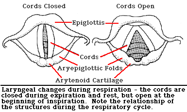
![]() The glottis is the area around the vocal cords. The subglottic area
is the area directly below the vocal cords leading into the trachea.
The changes in the larynx during the respiratory cycle are also
important to note.
The glottis is the area around the vocal cords. The subglottic area
is the area directly below the vocal cords leading into the trachea.
The changes in the larynx during the respiratory cycle are also
important to note.
![]() The cords are closed during the end of the expiratory phase and rest,
and they open at the beginning of the inspiratory phase. The
narrowest part of the adult airway is the vocal cords, but, in
children, the narrowest part is the cricoid cartilage located in the
subglottic area of the larynx.
The cords are closed during the end of the expiratory phase and rest,
and they open at the beginning of the inspiratory phase. The
narrowest part of the adult airway is the vocal cords, but, in
children, the narrowest part is the cricoid cartilage located in the
subglottic area of the larynx.
The trachea is a cylindrical structure formed by 16-20 U-shaped cartilaginous rings and a muscular/cartilaginous part that completes the tube. There is also some change in the shape of the trachea during inspiration and expiration.
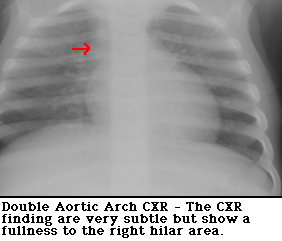
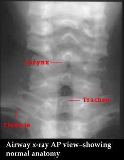
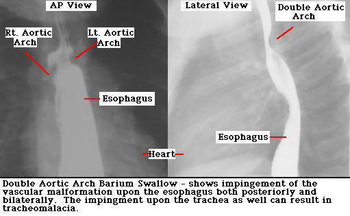
Imaging of the respiratory tract is common. Modalities include
plain chest x-ray,
![]() endolateral neck x-ray,
endolateral neck x-ray,
![]()
![]() barium swallow,
barium swallow,
![]() CT scan
CT scan ![]() ,
and cine CT
,
and cine CT ![]() .
.
Next Page | Previous Page | Title Page
Additional pediatric resources: GeneralPediatrics.com | PediatricEducation.org | SearchingPediatrics.com
Please send us comments by filling out our Comment Form.
All contents copyright © 1992-2024 Donna M. D'Alessandro, M.D. and Michael P. D'Alessandro, M.D. and the authors. All rights reserved.
"Virtual Pediatric Hospital", the Virtual Pediatric Hospital logo, and "A digital library of pediatric information" are all Trademarks of Donna M. D'Alessandro, M.D. and Michael P. D'Alessandro, M.D.
Virtual Pediatric Hospital is funded in whole by Donna M. D'Alessandro, M.D. and Michael P. D'Alessandro, M.D. Advertising is not accepted.
Your personal information remains confidential and is not sold, leased, or given to any third party be they reliable or not.
The information contained in Virtual Pediatric Hospital is not a substitute for the medical care and advice of your physician. There may be variations in treatment that your physician may recommend based on individual facts and circumstances.
URL: http://www.virtualpediatrichospital.org/